Hip And Leg Bone Diagram : Hip Anatomy Femur And Pelvis Bones That Make Up The Hip Joint And Acetabulum Smith Nephew Us Patient / Want to learn more about it?
byAdmin-
0
Hip And Leg Bone Diagram : Hip Anatomy Femur And Pelvis Bones That Make Up The Hip Joint And Acetabulum Smith Nephew Us Patient / Want to learn more about it?. The ilium bone forms the superior portion of the os coxa, the ischium bone the lower posterior portion, and the pubic bone (pubis) the lower anterior portion. Here's a diagram to sum up. The bones of the leg are the femur, tibia, fibula and patella. When you stand or walk, all the weight of your upper body rests on them. The knee joint is the largest joint in the body and is primarily a hinge joint, although some sliding and rotation occur.
Your leg bones are the longest and strongest bones in your body. Diagram of blood and nerve supply to bone. It is the most complete reference of human anatomy available on web, ipad, iphone and android devices. The pelvis and the femur (the thighbone). The foot bones shown in this diagram are the talus, navicular, cuneiform, cuboid, metatarsals and calcaneus.
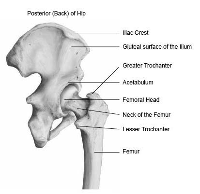
Hip Anatomy Pictures Function Problems Treatment from www.healthpages.org Hip bone diagram wiring diagram. Your leg bones are the longest and strongest bones in your body. Normally, a smooth cushion of shiny white hyaline (or articular) gluteus medius and minimus are the main abductors of the hip —that is, they move the leg away from the midline of the body (using the spine as a midline. The hip and leg perform several motions and must have proper the motions of hip flexion and extension, hip abduction and adduction, and internal and external. On top of that layer of muscle is the iliotibial band, which starts at the brim of your pelvis outside the hip joint and runs down your leg. Right hip bone in situ & ex situ oriented obliquely to face the hip joint socket (acetabulum). Download this free vector about diagram showing the hip bone treatment, and discover more than 15 million professional graphic resources on freepik. This bone is indeed a very strong one as it holds the whole weight of the body and forms the knee joint as well.
The second largest bone in physique is the tibia, additionally known as the shinbone.
High resolution textures and displacement included. Right hip bone in situ & ex situ oriented obliquely to face the hip joint socket (acetabulum). The head of your femur fits into your hip socket and the bottom end connects to your knee. When you stand or walk, all the weight of your upper body rests on them. The hip joint gives the leg an incredible range of motion while still providing support to the body's weight. Ct, mri, radiographs, anatomic diagrams and nuclear images. Download this free vector about diagram showing the hip bone treatment, and discover more than 15 million professional graphic resources on freepik. Explore over 6700 anatomic structures and more than 670 000 translated medical labels. The bones involved in it, however, are only the femur and the tibia, although the smaller bone of the leg, the fibula, is carried along in the movements of flexion, extension, and slight rotation that this joint. Normally, a smooth cushion of shiny white hyaline (or articular) gluteus medius and minimus are the main abductors of the hip —that is, they move the leg away from the midline of the body (using the spine as a midline. Fibula and tibia, ankle and foot. 3d illustration of human body hips stock illustration illustration. Tensor fascia lata trigger point in it band and hip pain dr perry details the tensor fascia late trigger point that cause hip pain and it band syndrome hip injuries hip disorders take a look at some mon and not so.
Hip muscle strains info florida orthopaedic institute. Hip bone diagram wiring diagram. Archeologists in belgium have discovered walls made. The bones of the leg are the femur, tibia, fibula and patella. The head of your femur fits into your hip socket and the bottom end connects to your knee.
Hip Hop Your Way To Health How To Support Hips As We Age from static.wixstatic.com Select from premium hip diagram of the highest quality. Download this free vector about diagram showing the hip bone treatment, and discover more than 15 million professional graphic resources on freepik. Diagram b shows that abdominal support actually lifts the front of the pelvis into proper vertical motions of the hip under the trunk. The pelvis and the femur (the thighbone). Ct, mri, radiographs, anatomic diagrams and nuclear images. Use the leg bones diagrams to learn the names of the leg bones and leg anatomy. Hip and thigh bones joints muscles kenhub. It joins the lower limb to the pelvic girdle.
Click and start learning now!
The foot bones shown in this diagram are the walls made of human skulls and leg bones uncovered next to belgian church | cbc radio. The bones involved in it, however, are only the femur and the tibia, although the smaller bone of the leg, the fibula, is carried along in the movements of flexion, extension, and slight rotation that this joint. Let's assume this figure is standing with the feet vertically aligned with the hip in an erect posture, you can place the pelvic bone (a narrower version of the head's egg), the this completes the basic, undifferentiated human proportions drawing tutorial. High resolution textures and displacement included. Hip and leg bone markings. The knee joint is the largest joint in the body and is primarily a hinge joint, although some sliding and rotation occur. Hip anatomy, function and common problems. Find the perfect hip diagram stock photos and editorial news pictures from getty images. Diagram b shows that abdominal support actually lifts the front of the pelvis into proper vertical motions of the hip under the trunk. Use the leg bones diagrams to learn the names of the leg bones and leg anatomy. Human body bone joint pains anatomy stock illustration 702398404. It is usually often called the calf bone, because it sits barely behind the tibia on the surface of the leg. Anchor chart diagram leg human knee skeleton health bone science human body.
Your leg bones are the longest and strongest bones in your body. Front view of the hip joint bones. The foot bones shown in this diagram are the walls made of human skulls and leg bones uncovered next to belgian church | cbc radio. The foot bones shown in this diagram are the talus, navicular, cuneiform, cuboid, metatarsals and calcaneus. These same nerves innervate the knee, which explains why pain can be referred to the knee from the hip and vice versa.
Hip Joint With Legs Stock Illustration Illustration Of Joint 46887320 from thumbs.dreamstime.com Hip muscle strains info florida orthopaedic institute. A diagram of the human skeleton. The hip joint gives the leg an incredible range of motion while still providing support to the body's weight. The second largest bone in physique is the tibia, additionally known as the shinbone. The pelvis and the femur (the thighbone). The two bones beneath your knee that make up your shin are. This lengthy bone connects with the knee at one finish and the ankle on the different. Hip and thigh bones joints muscles kenhub.
Explore over 6700 anatomic structures and more than 670 000 translated medical labels.
Hip and thigh bones joints muscles kenhub. The hip joint gives the leg an incredible range of motion while still providing support to the body's weight. Archeologists in belgium have discovered walls made. Fibula and tibia, ankle and foot. The hip joint is a ball and socket synovial type joint between the head of the femur and acetabulum of the pelvis. Find the perfect hip diagram stock photos and editorial news pictures from getty images. A diagram of the human skeleton. Front view of the hip joint bones. Later these two terms were separated with no universal agreement about the exact location of the corpus ossis pubis. Here's a diagram to sum up. Hip anatomy, function and common problems. Download this free vector about diagram showing the hip bone treatment, and discover more than 15 million professional graphic resources on freepik. Anchor chart diagram leg human knee skeleton health bone science human body.
High resolution textures and displacement included leg bone diagram. These same nerves innervate the knee, which explains why pain can be referred to the knee from the hip and vice versa.